We specialise in stock science images, which are taken using a scanning electron microscope (SEM). This technology allows us to produce detailed scanning electron micrographs of things that cannot normally be seen, such as blood cells, chromosomes, and microscopic flora and fauna.
Electron micrograph images are produced by the microscope in shades of grey. These microscope images have to be colourised to produce the coloured micrographs that we offer here at PSmicrographs. When scanning electron microscopes first appeared during the early 1960s, the images wowed the world, but they were all in black and white; it wasn’t until software was introduced to colour these micrographs that they truly came to life.
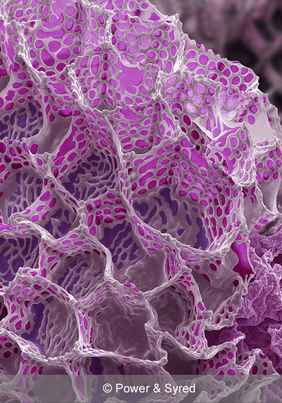
Foxglove Seed
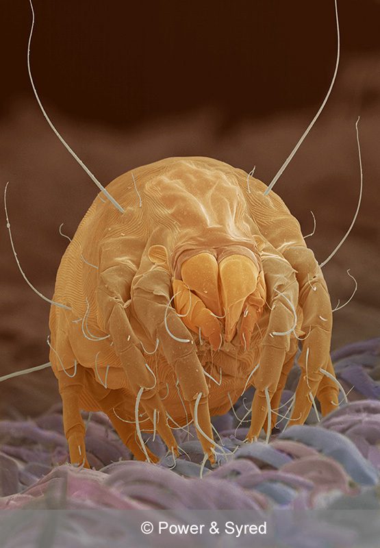
House Dust Mite (Dermatophagoides pteronyssinus)
Creating scanning electron micrographs
Many people do not realize how much work goes into producing our stock science images – obtaining a suitable sample to use on an SEM is just the start of the process! Each sample has to be cleaned and treated depending on its nature (for example, soft tissue would be treated differently to a rock sample); delicate samples like soft tissue can take hours and sometimes days to prepare. It is never a cut-and-dried procedure, and it is quite common for samples go wrong, which means that the whole procedure has to be re-started with new samples.
The Scanning Electron Microscope (SEM)
Most samples have to be coated in a conductive material to stop the specimen charging in the SEM vacuum chamber, where the sample is bombarded with electrons. This coating involves the use of a sputter coater, which covers the specimen in a fine coat of gold, platinum, palladium or some other suitable conductive material. If the specimen is of soft tissue (like a small insect, a piece of plant material, or even a human sample of blood or perhaps chromosomes), the sample has to be dried, either with chemicals or with a critical point dryer, a machine that takes all the moisture out of the plant, insect or blood sample. If the sample is not dried then it will vent within the sputter coater and the vacuum chamber, causing numerous problems with the sample and the equipment.
Preparingthe specimen
Once the specimen is dried sufficiently it can be coated, but the drying process is sometimes not effective, in which case the sample will distort or collapse under vacuum. This is very often the case with insects such as dust mites, fly larvae, mosquito pupa and particularly tough skinned insects. Plant material invariably has a distortion problem no matter how carefully the specimen is prepared.
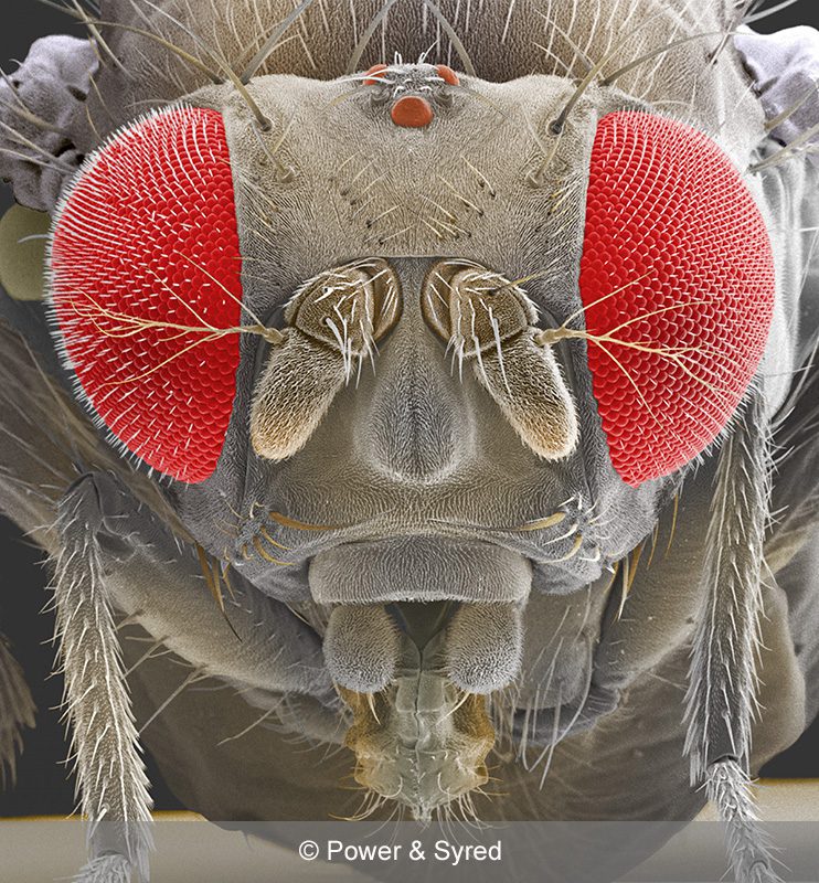
Fruit fly head (Drosophila melanogaster)
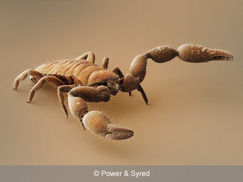
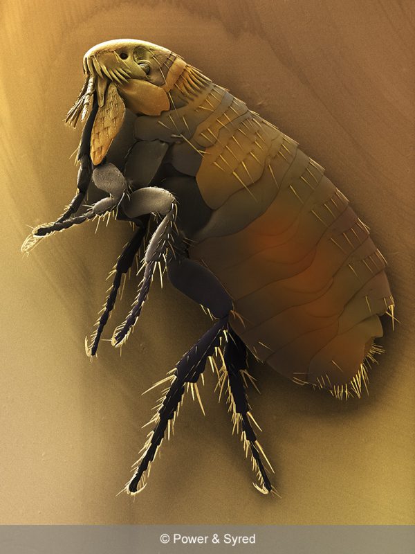
Dog flea (Ctenocephalides canis)
Setting out microscopic animals
With respect to microscopic animals (such as fruit flies, small spiders, and weevils), these creatures have to be set out before drying. Their legs must be arranged as if they are alive, and this can be a delicate and tricky operation, especially when fine antenna and many legs are involved. Losing a leg or an antenna is a common occurrence, so more than one sample will be set out at a time to allow for losses. Taking a dead fly from the kitchen windowsill is not an option; the insect will never provide a lifelike sample if it looks dead before it is prepared.